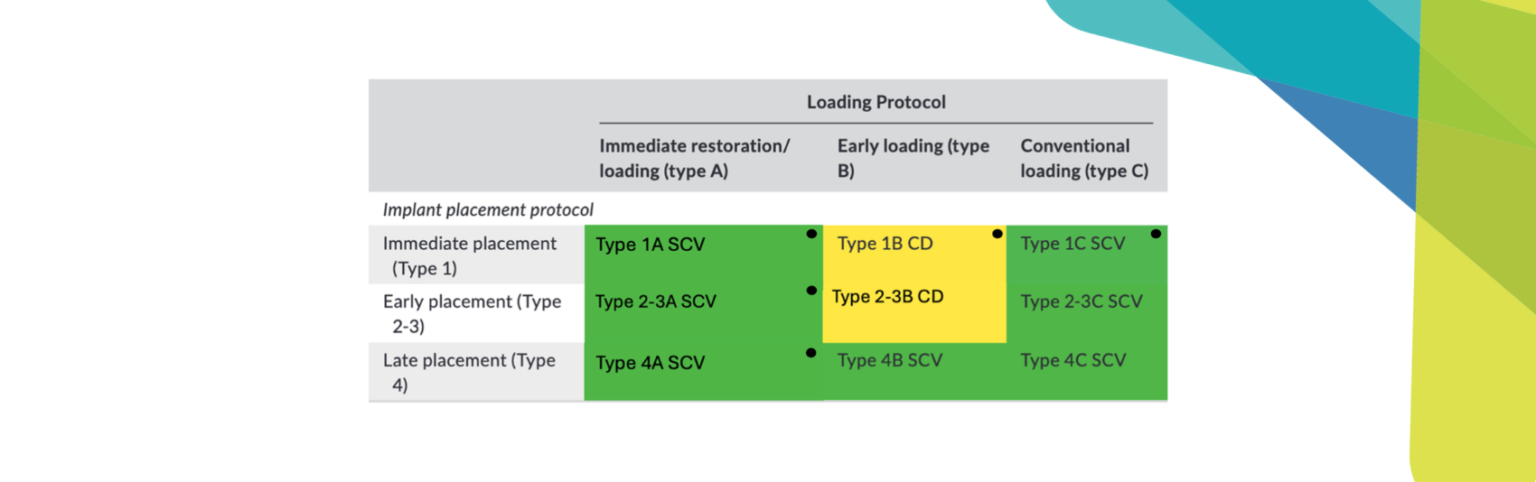
The essential checklist for long-term implant success
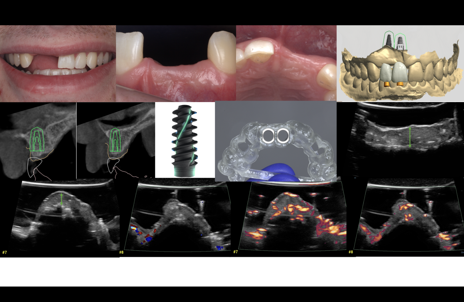
In daily practice, we mostly rely on what we can see clinically or what appears calcified on radiographs. The true “black box” of implant dentistry, however, is the softtissue interface. Conventional tools often fail to capture the early biological dynamics of softtissue integration, subtle inflammatory changes, or their real-time resolution. Intraoral ultrasonography (IUS) offers a noninvasive, radiation-free window into peri-implant healing, allowing quantitative assessment of mucosal thickness and vascularity in real time.