Interdental papillae dimensions and patient satisfaction
The presence and size of interdental papillae are decisive for patient satisfaction and can significantly impact treatment outcomes (Karateew, 2017). For instance, the lack of papillae (black space/triangle) between the maxillary central incisors negatively impacts the perception of facial esthetics (Al-Lahham et al., 2021). In addition, the size of the black space can determine the extent of this negative effect (Al-Lahham et al., 2021).
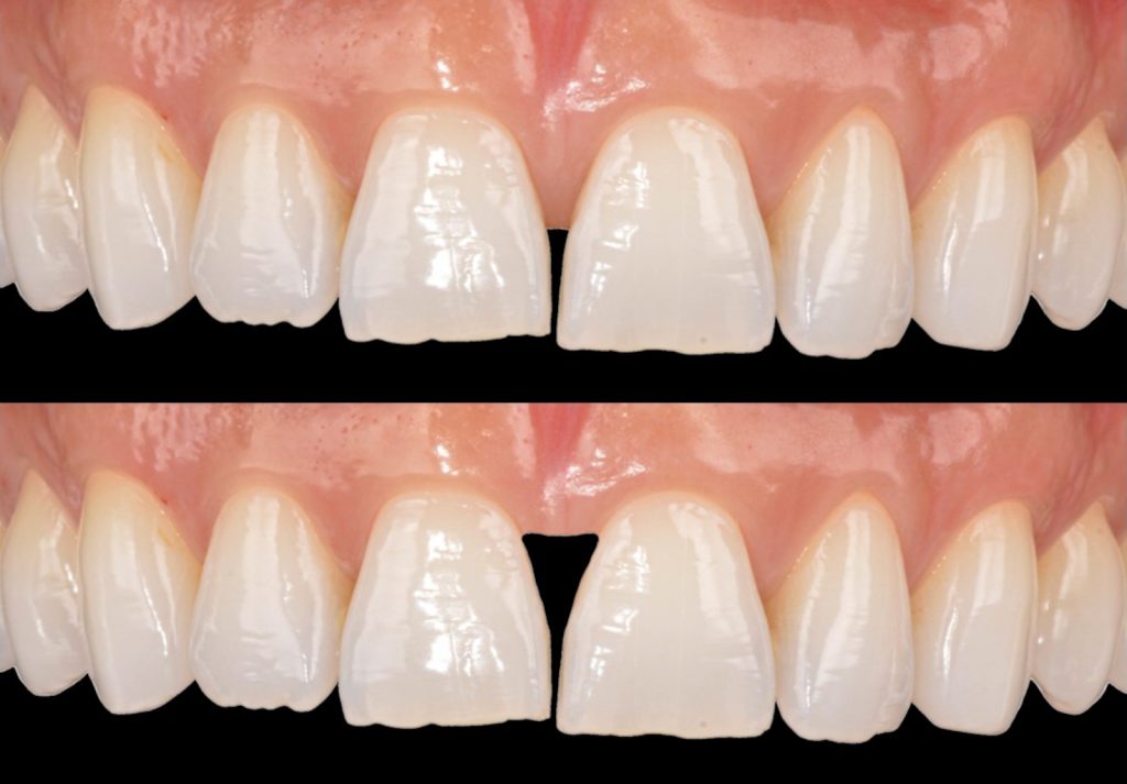
Fig. 1: An increase in the size of the black space can bring more attention to the lack of soft tissue (the original photo is a courtesy from Dr. med. dent. MSc PhD. Gabriela Sabatini and the interdental papillae was edited to illustrate the black space)
The optimal papillae design can be achieved when the volume exceeds that of the buccal aspect of the teeth, and the length is approximately half that of the teeth (Chu et al., 2009). Bilateral variations in papillae length within the 2- to 3-mm range are considered esthetically acceptable (LaVacca et al., 2005). However, asymmetrical deviations greater than 2 mm in papillae length are considered unattractive (Yu et al., 2015). Additionally, papillae height tends to decrease by 0.012 mm each year with increasing age, and complete papillae filling is associated with an interproximal gingival tissue thickness of ≥ 1.5 mm (Chow et al., 2010).
Several factors can influence the papillae dimensions, including the interproximal bone crest of the adjacent tooth and the presence of an interproximal contact point (Nisapakultorn et al., 2010; Tarnow et al., 1992; Ishikawa et al., 2010). The papillae volume has also been correlated with crown proportions and transgingival probing (Shao et al., 2018). In addition, reduced papillae volume was more frequent in average-scalloped teeth and thin-scalloped soft tissue phenotypes (Shao et al., 2018).
Understanding soft tissue
Ineffective diagnosis and treatment planning can result in multiple unsuccessful interventions, which may fail to meet patient expectations (Parize et al., 2022). Therefore, achieving optimal treatment outcomes requires a comprehensive approach to soft tissue that considers the biological morphology, prosthetic requirements, and esthetic perceptions. The lecture entitled “Biological Basics in Soft Tissue Management” by Prof Dr Johannes Kleinheinz, available at the ITI Academy, supplies the necessary background for establishing individualized and appropriate soft tissue management that takes the anatomical, biological, and physiologic aspects of soft tissue into consideration.
Biological Basics in Soft Tissue Management
This ITI Academy lecture discusses the anatomical, biologic, and physiologic aspects of the soft tissue, which play a decisive role during the initial assessment of the patient’s situation and the resulting treatment planning.
Method overview
There are an increasing number of reports on the use of digital tools to assess periodontal and peri-implant tissue dimensions (Tavelli et al., 2021; Kuralt et al., 2022). Despite the different workflows available for the detailed assessment of papillae dimensions and stability over time, this article focuses on two three-dimensional (3D) methods: 1) Standard tessellation file (STL) and 2) STL file combined with cone beam computed tomography (CBCT). In addition, the benefits and drawbacks of each method are presented, along with a step-by-step workflow.
Standard tessellation files (STL)
STL files can be obtained directly from intraoral scanners or by digitalizing conventional impressions of cast models. STL files associated with metrology software have evolved from designing restorations to assessing volume changes of hard and soft tissues in various clinical scenarios (Tavelli et al., 2021). This method involves assessing changes in volume by overlaying three-dimensional (3D) images captured at various time intervals.
Using an intraoral scanner to assess papillae tissue represents a quick, non-invasive (lack of ionizing radiation), and comfortable (lack of anesthesia and bone sounding with needles or endodontic files) method that allows clear, real-time communication with clinicians and patients (Dineen & Brennand Roper, 2020). Compared to analog impression materials, improved patient comfort and lack of compression and deformation of soft tissues are beneficial for evaluating postoperative (wound healing) and long-term patient monitoring (tissue stability) (Dineen & Brennand Roper, 2020; Zhang et al., 2021). Although this method lacks information on bone anatomy (Couso-Queiruga et al., 2021), it can provide information regarding the need for augmentation procedures (Mancini et al., 2023) and provide insights regarding alveolar bone resorption after socket preservation procedures due to the volumetric modification of soft tissues (Ivanova et al., 2019). In addition, gingival recession (Fageeh et al., 2019) and keratinized gingiva width (Lim et al., 2021; Lee et al., 2020) can be more accurately measured with intraoral scanned data than with conventional techniques. However, repeated scans are needed, the accuracy of which can be influenced by several factors (Revilla-León et al., 2023a, 2023b), and the selected matching regions for data alignment can influence the measurements obtained (Kuralt & Fidler, 2021). In addition, the experience and learning curve associated with software should also be considered in terms of workflow accuracy (Tavelli et al., 2021).
The use of reverse engineering software such as ZEISS Inspect (previously known as GOM Inspect) and Geomagic (3D System) has been extensively reported (Kuralt et al., 2022). Despite being established as reliable software, the high acquisition price and complex steps are obstacles to implementing this workflow in clinical practice (Kuralt et al., 2022). The necessity of multiple specialized software programs has also been reported (Kuralt et al., 2022). Therefore, optimal software should prioritize user-friendly interfaces, open-source availability and incorporate a full range of evaluation tools (Kuralt et al., 2022).
Medit Link (Medit) is an open-source collaboration tool that allows improved communication between clinicians, patients, and third parties by integrating computer-aided design (CAD) software, dashboards, cloud storage and scan data synchronization. In addition, deviation measurement tools, which were previously restricted to reverse engineering software, are also available with a user-friendly interface. Similar deviation measurements were also reported when Medit Link was compared to other established metrology software (Cakmak et al., 2022; Dede et al., 2023).
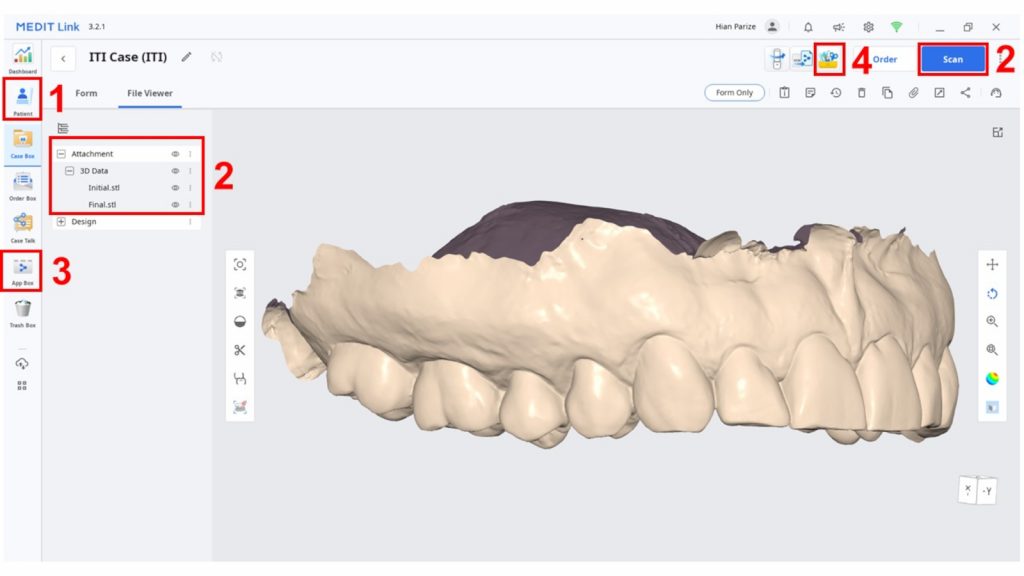
Fig. 2: Medit Link steps 1 to 4. Patient case creation (1), dataset installation (2), app installation (3) and launching (4) 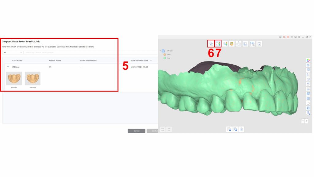
Fig. 3: Medit Link steps 5 to 7. The data are imported to app (5), alignment (6) is performed, and deviation mode (7) is selected 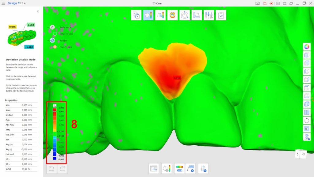
Fig. 4: Medit Link step 8. The color label was adjusted according to case-specific clinical settings 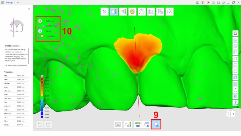
Fig. 5: Medit Link steps 9 and 10. Cross-sections (9) and (10) show the model visibility functions 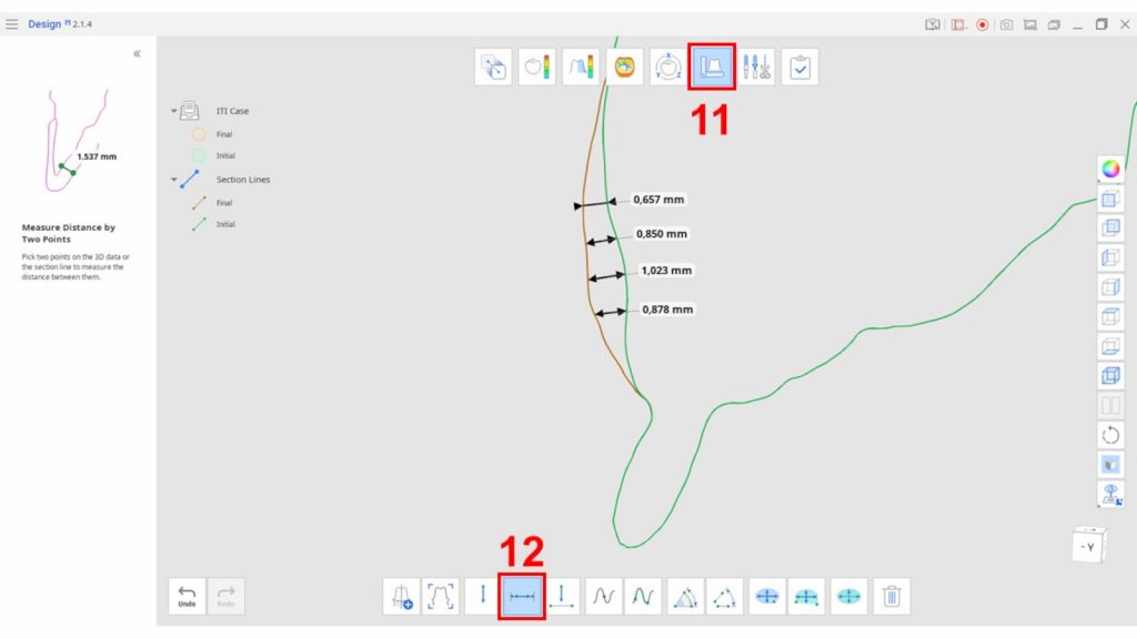
Fig. 6: Medit Link steps 11 (measurement mode) and 12 (measure distance by two points) 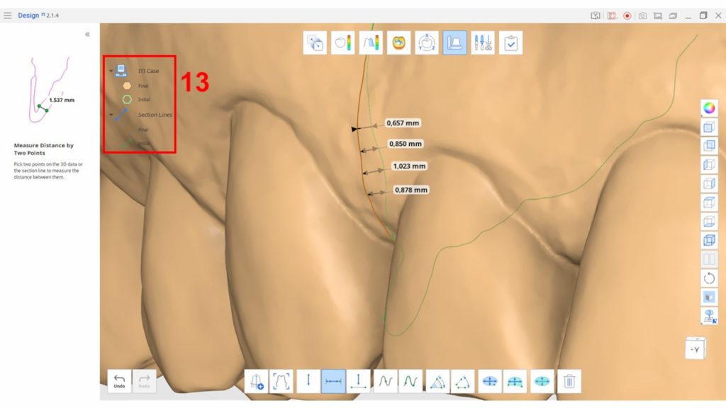
Fig. 7: Medit Link step 13. Model appearance settings 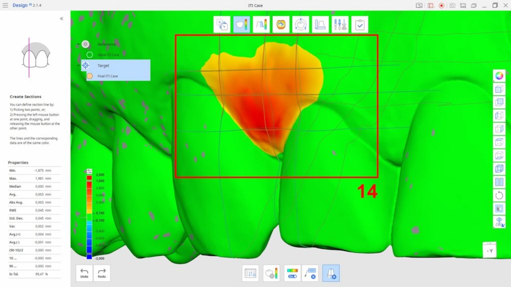
Fig. 8: Medit Link step 14. Multiple cross-sections 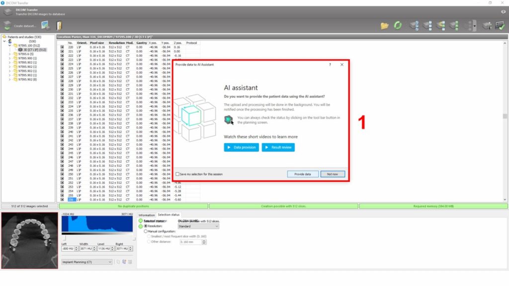
Fig. 9: coDiagnostiX step 1. Patient dataset creation and hard tissue segmentation 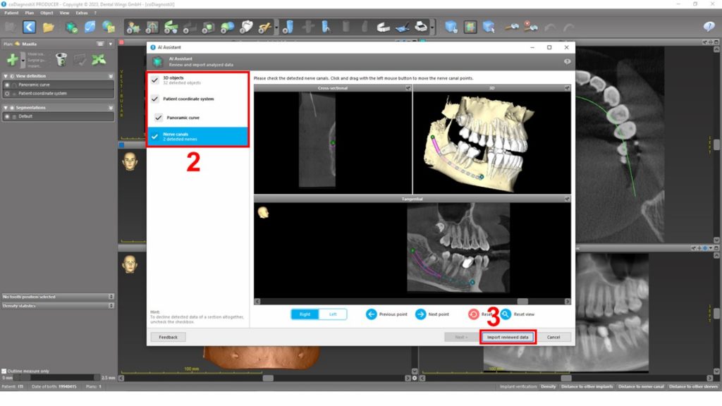
Fig. 10: coDiagnostiX steps 2 and 3. The AI-segmented data were reviewed and imported 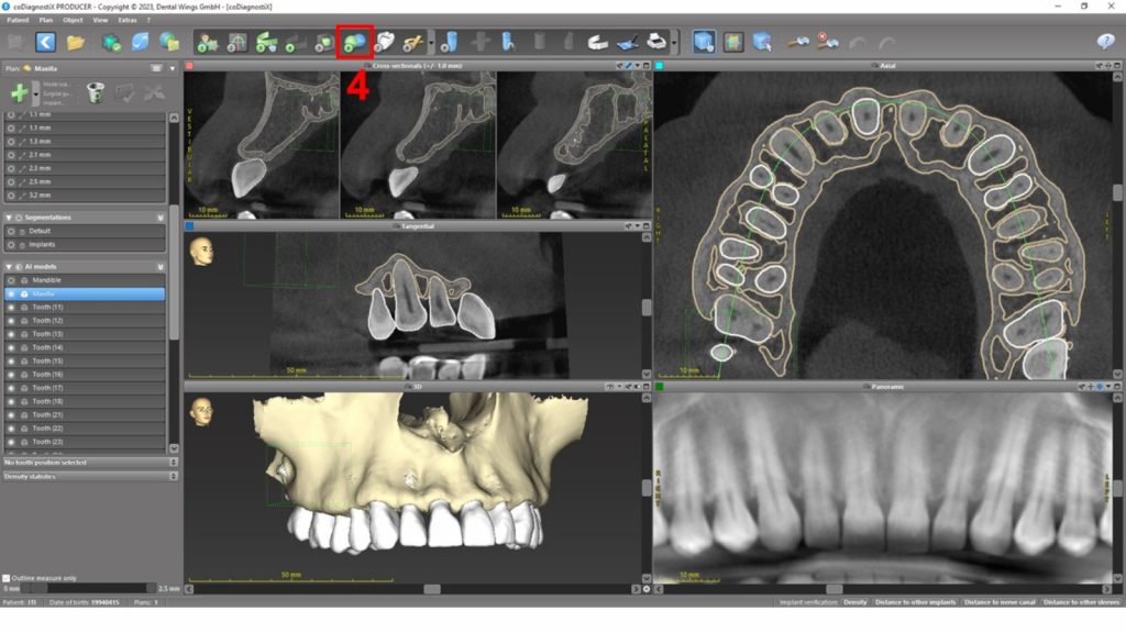
Fig. 11: coDiagnostiX step 4. IOS data import and AI-assisted alignment 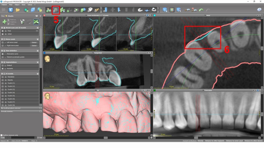
Fig. 12: coDiagnostiX steps 5 and 6. 2D reference lines and area of interest adjustment 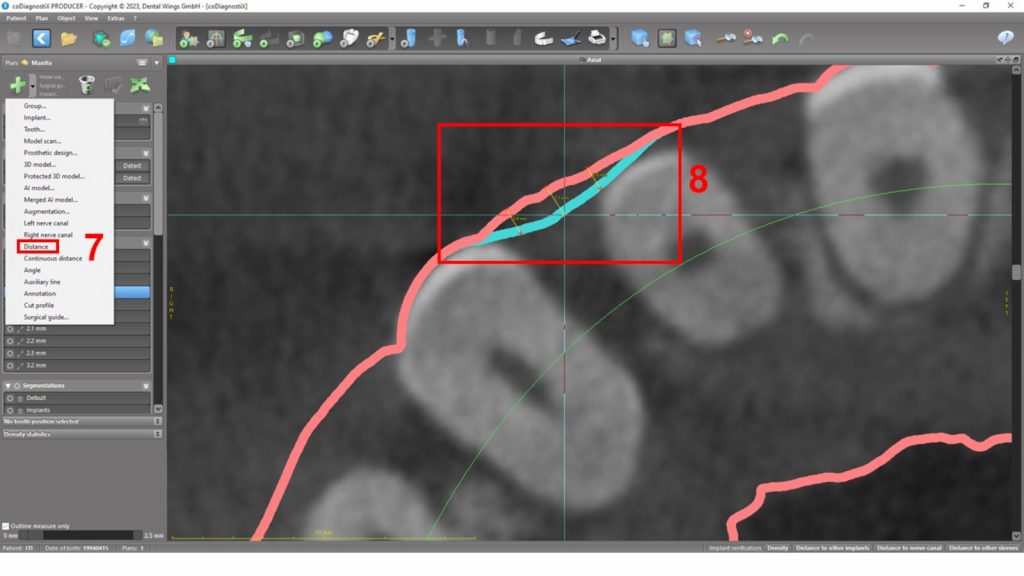
Fig. 13: coDiagnostiX steps 7 and 8. Addition of linear measurements 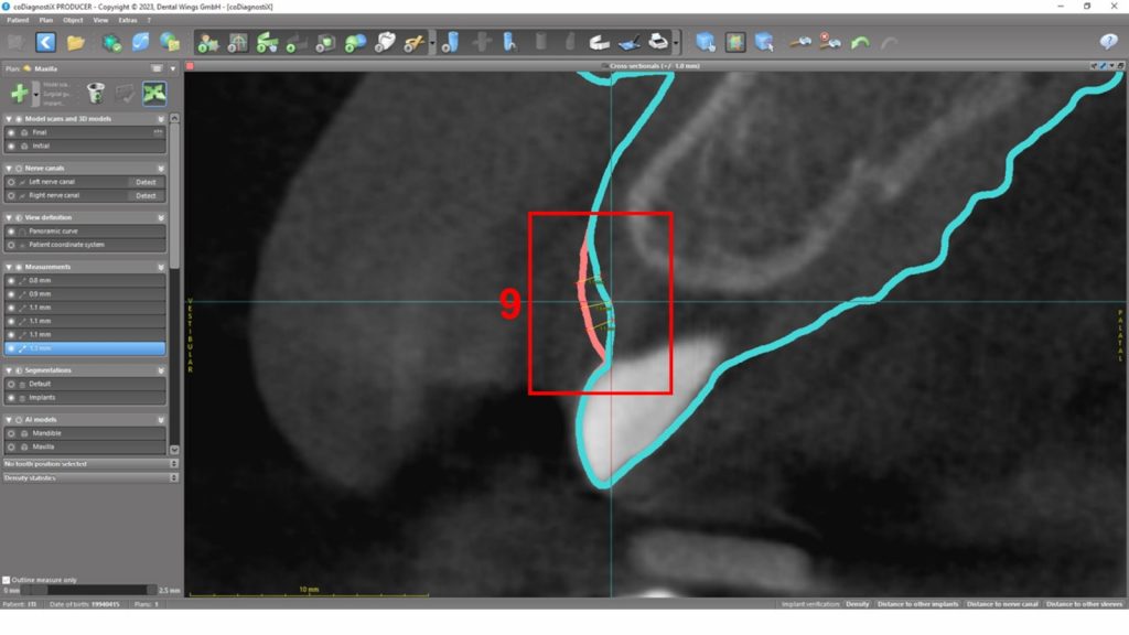
Fig. 14: coDiagnostiX step 9. Linear measurements on a cross-sectional view 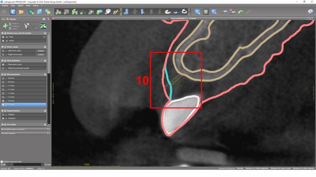
Fig. 15: coDiagnostiX step 10. Varying model visibility allows improved visibility of hard-to-soft tissue measurements 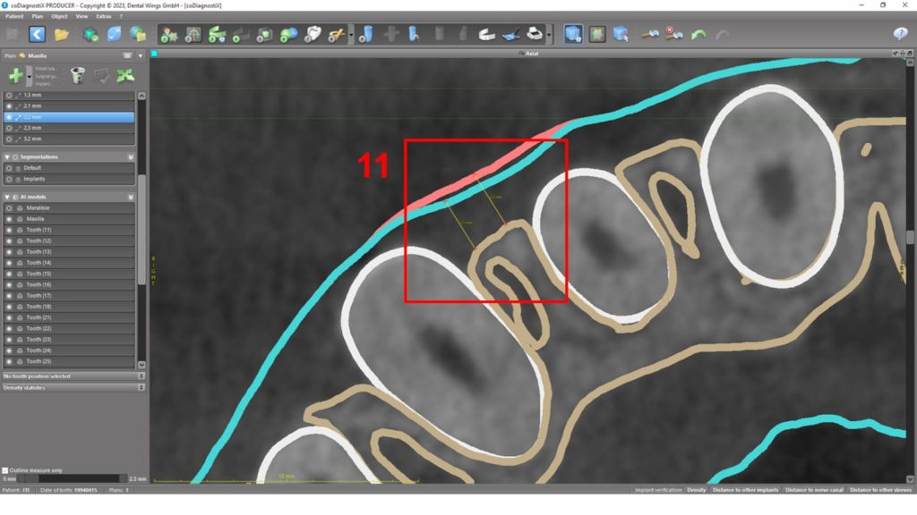
Fig. 16: coDiagnostiX step 11. Axial view and hard-to-soft tissue measurements
STL file + cone-beam computed tomography (CBCT)
Despite the use of several classification systems for the assessment of papillae loss (such as the Nordland and Tarnow Classification, Papillae Presence Index Papillae Index Score, and Papillae Fill Index), there is still a lack of classification systems using recent digital tools that allow for 3D volume assessment over time. In addition, the distance between the interproximal bone crest and interproximal tooth contact point can strongly influence the papillae height and volume (Nisapakultorn et al., 2010; Tarnow et al., 1992; Ishikawa et al., 2010).
CBCT is an imaging technique that allows for the detailed 3D assessment of mineralized tissues. However, it has lower contrast to soft tissues, leading to inaccurate contouring of periodontal and peri-implant tissues (Korostoff et al.,, 2018). Therefore, cotton or lip/tongue retractors were introduced to enable proper, refined visualization of soft tissues, allowing measurement of both hard and soft tissues (Januário et al., 2011). Later, the alternative technique of associating intraoral scans was introduced (Kim et al., 2016). This allowed a detailed assessment of the bone anatomy and the need for hard and soft tissue augmentation (Couso-Queiruga et al., 2021; Mancini et al., 2023; Ko et al., 2021). For instance, previous studies reported high agreement between this technique and histological assessment of soft tissue thickness, providing a detailed assessment of the periodontal phenotype (Ferry et al., 2022). However, the artifacts induced by metal and ceramic materials can impact the visualization and measurement of soft tissues (Couso-Queiruga et al., 2023).
The assessment of intraoral anatomical structures using CBCT must respect the ALARA (as low as reasonably achievable)/ALADA (as low as diagnostically acceptable) principles (Jaju & Jaju, 2015). Therefore, the presence of ionizing radiation represents a drawback to this technique, and case-specific indications should be addressed with caution. For instance, variations in DICOM resolution can influence radiation exposure, requiring an evaluation of the risk-benefit ratio (Ferry et al., 2022). Conversely, recent advancements in magnetic resonance imaging (MRI) of mineralized tissues (e.g., bone and teeth) represent an alternative clinical workflow (Flügge et al., 2023; Flügge et al., 2023; Greiser et al., 2024).
The combination of STL and CBCT provides a comprehensive and precise assessment of periodontal and peri-implant tissues by integrating the strengths of both digital methods. STL files allow for non-invasive, radiation-free, and highly detailed surface scans, enabling real-time monitoring of soft tissue changes, gingival recession, and keratinized gingiva width with high accuracy. Meanwhile, CBCT provides essential insights into underlying bone anatomy, aiding in the evaluation of interproximal bone crest levels and the need for augmentation procedures. By merging these modalities, clinicians can achieve a more holistic understanding of the periodontal phenotype, facilitating personalized treatment planning while minimizing radiation exposure through case-specific indications. This integrated approach enhances diagnostic accuracy, treatment predictability, and long-term patient outcomes by optimizing both soft and hard tissue evaluations. However, the integration of these methods requires careful consideration of factors such as ionizing radiation exposure in CBCT and the learning curve associated with advanced software tools.
Conclusion
The intraoral scanner and cone-beam computed tomography systems can be applied with different techniques to perform 3D assessments of interdental papillae.