Introduction
In the second part of the blog, we had discussed about the lateral window and transcrestal approach for sinus floor elevation along with complications. In the third part of this blog article, we will be covering the alternatives/advancements to these two techniques which were proposed in order to minimize the potential complications, and improve patient perception.
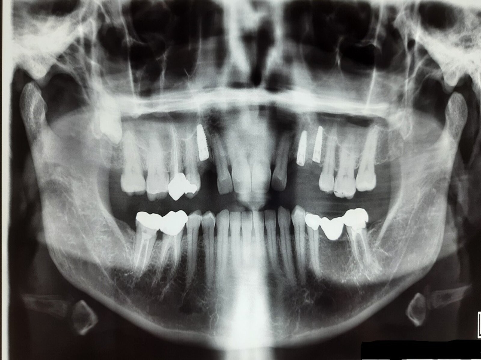
Selection of implant in the maxillary posterior segment
The following parameters should be assessed prior to selection of an implant at the posterior site with planned sinus floor elevation procedure:
- Implant diameter
- Implant shape
- Implant surface
Implant diameter and shape primarily influence the primary stability of the implant which is critical for osseointegration. Tapered implant shapes, especially if shorter implants are placed (Barikani H et al. 2014), and wider diameter cylindrical implants help to optimize primary stability (Lozano-Carrascal et al. 2016).
The implant surface determines the quality and dynamics of the bone formed over the implant during the initial healing period. This influences the quality of anchorage provided and the interval required before loading the implant. Microrough surface implants are preferred at sites where sinus floor elevation is performed owing to faster bone apposition over the implant surface and higher survival rates (Buser et al. 2017).
Advances/Alternatives
Short implants have been considered as an alternative in the posterior maxilla owing to their less invasive nature. Short (6-mm) implants have been used to circumvent sinus floor elevation and grafting. Investigations have been conducted by multiple researchers regarding the predictability and complications in relation to use of short implants.
Lemos et al. (2016) considered short implants to be a predictable treatment alternative for the posterior jaw. However, they stated that implants under 8 mm in length (4-7 mm) present with a greater risk of failure compared to standard implants, and should be used with care. Papaspyridakos et al. (2018) stated that shorter implants (≤6 mm) presented with higher variability and lower predictability in survival rates compared to longer implants (>6 mm) after 1-5 years in function. The mean survival rate for shorter implants was found to be 96% (range: 86.7%-100%) in comparison to 98% (range 95%-100%) for longer implants. Based on the evidence generated, short implants of ≤6 mm length should be selected after careful consideration since they present with a greater risk of failure.
Cruz et al. (2018) stated that short implants are an effective substitute for sinus augmentation and long implants due to fewer biological complications, and comparable marginal bone loss and survival rates. However, higher rates of prosthetic complications were noted with the use of short implants.
It is important to consider whether the implants reported in the studies were single standing or multiple adjacent short implants that were splinted. Studies have shown that single-standing implants present with a greater failure rate compared to multiple splinted short implants (Felice et al. 2014; Rossi et al. 2016).Ultra-short implants of less than 4 mm in length have been used in exceptional circumstances. However, they should always be splinted with a longer implant since the crown implant ratio is greater than 2 and the presence of any parafunctional habits might trigger crestal bone loss (Buser et al. 2017).
Piezoelectric surgery
Piezoelectric bone surgery was developed to overcome the limitations of traditionally instrumented oral bone surgery (Vercellotti et al. 2000). The concept of piezoelectric surgery is centered on the piezoelectric effect, where certain ceramics and crystals deform when an electric current passes through them, producing oscillations at an ultrasonic frequency. The vibrations obtained are then amplified and directed to a vibration tip, which when applied with light pressure to a hard bony tissue produces a cavitation phenomenon, an effect seen entirely in mineralized tissues (Fig 1& 2). The cavitation effect induces hydropneumatic pressure of the saline irrigant that helps to elevate the sinus membrane without trauma or peforation.
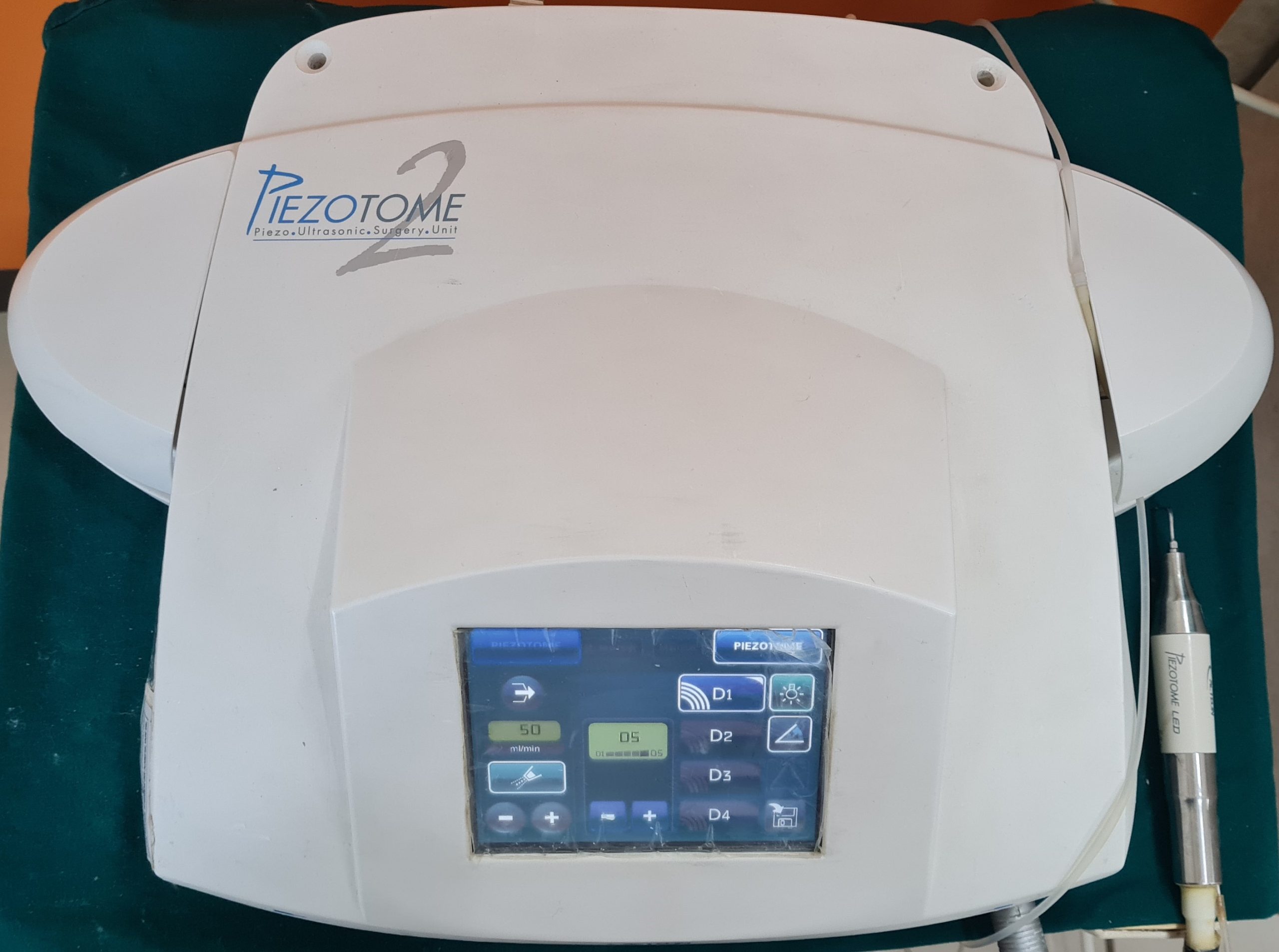
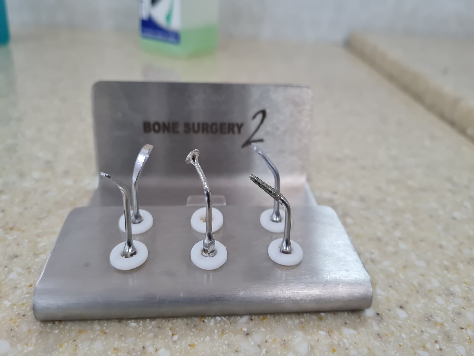
It must be noted that morphological characteristics of the piezosurgical osteotomies varied depending on the piezosurgical unit and tip used (Bauer & Romanos 2014). The active tip of the piezosurgical device is small, which results in precise and selective cutting , and enhanced safety for the patient (Stubinger et al. 2008). The active tip does not damage any soft tissues, for instance neurological tissues, since it vibrates at 25-30KHz frequency while soft tissues require frequency above 50KHz. An important benefit of piezosurgery is the relatively blood-free surgical site compared with the use of conventional microsaws, which ensures good visibility during the procedure (Stubinger et al. 2008), and contributes towards improved healing conditions. The constant irrigation during the course of peizosurgery also helped to reduce any thermal damage and reduce the risk of bone necrosis during implant site preparation.
The sinus lift procedure includes the removal of a bony window of the anterior sinus maxillary wall followed by sinus membrane elevation. The prevalence of sinus membrane perforation with rotary instrumentation varies between 5% and 56% whereas a 5% reduction in membrane perforation using piezoelectric surgery was reported (Kasabah et al. 2003; van den Bergh et al. 1998, Vercellotti et al. 2000, Seoane et al. 2013; Stacchi et al. 2015, Stacchi et al. 2017). After sinus membrane elevation, the implant site may be prepared at the same appointment using piezoelectric instrumentation and a reduction in inflammatory cells and increased osteogenic activity around implants placed by a piezoelectric ultrasound device in comparison with other systems was documented (Preti et al. 2007, Robiony et al. 2007). A minor drawback in regard to the use of piezoelectric equipment would be the extended surgical time in relation to conventional rotary instruments (Stacchi et al.2020).
Hydraulic sinus lift technique
In 2010, the hydraulic sinus lift, a new method, was proposed in which the antral mucosa is detached and the sub-antral space simultaneously filled by applying hydraulic pressure on a graft material of a pasty consistency (Andreasi & Lopez 2010).
The device created for this purpose comprises three components: a titanium syringe equipped with a micrometric piston to assemble single-use plastic syringes of various volumes, a dispenser in threaded surgical steel available in different forms and measurements, and a surgical steel needle. The single-use syringes can be pre-loaded with the desired amount of graft material, or it is possible to directly use the syringe containing the graft material as provided by the manufacturer.The hydraulic sinus lift technique has been shown to be advantageous with a short learning curve, minimal invasiveness, and greater precision. They have also documented the relatively low risk of membrane perforation utilizing this technique and considered it safe for performing sinus floor elevation (Andreaasi et al. 2017; Kim et al. 2012).
Balloon elevation technique
Minimally invasive antral membrane balloon elevation (MIAMBE) is a modification of the transcrestal technique, whereby the antral membrane elevation is performed through the osteotomy site (3.5 mm) using a specially designed balloon.The MIAMBE device consists of a stainless steel tube, 3 mm in diameter, connected to an inflation syringe at its proximal end, and an embedded single use silicone balloon at its distal end. Diluted contrast fluid is used to inflate the balloon which pushes up the Schneiderian membrane, and creates the desired space for implant placement (Kfir et al. 2007; Kfir et al. 2009A; Kfir et al. 2009B; Dhandapani et al. 2016; Asmael 2018).Once the desired lift is obtained, the balloon remains inflated in the sinus for five minutes to reduce the elasticity of the sinus membrane, following which it is deflated and removed. Bone graft material is then placed, followed by implant placement at the osteotomy site.
Minimally invasive antral membrane balloon elevation combines the advantages of both the lateral window approach and transalveolar approach, producing ≥10 mm of bone gained in a minimally invasive manner. Utilizing this technique, the membrane perforation rate was relatively low at 6.76%, and sinus augmentation success rate averaged 91.6% (Asmael 2018).
Huwais (2018) developed a new implant site/osteotomy preparation technique utilizing non-subtractive drilling in which the bone density increases as the implant site is prepared, referred to as osseodensification (Huwais 2018).
This surgical technique uses specially designed fluted burs (DENSAH burs) that help densify the bone as the osteotomy site is prepared. Conventional or standard drills remove bone during the osteotomy, whereas the DENSAH burs ensure bone preservation and condensation by compaction, thereby, increasing the bone density in the peri-implant area and improving mechanical stability (Lipton et al. 2015; Trisi et al. 2016). This technique, though introduced for horizontal augmentation in narrow alveolar ridges, can also be used to gain vertical height with a transcrestal approach. According to the inventor’s DENSAH protocol, if additional lift floor elevation beyond 3 mm is required, an allograft material can be placed and gently pushed into the sinus space to achieve an additional 2 mm in height. Following the lift, the implant is placed into the site with the appropriate torque wrench (Huwais 2018).
Conclusion
Sinus floor elevation is a predictable surgical procedure. Apart from the traditional techniques, alternatives have been proposed in order to reduce compications an improve patient perception. The clinician needs to be aware of such alternatives in terms of instrumentation or armamentarium to remain updated and enhance his/her clinical skills for the benefit of dentalcommunity and patients.
Read Part 2: Overview of sinus floor elevation procedure in implant dentistry – Part 2
Discover more about this topic: