Introduction
We have seen many advances in dental implant designs and surface technologies in recent years. For some time, however, the prosthodontic management and components have been based on a conventional analog laboratory pathway utilizing burn-out and metal castings that deliver a wide spectrum in accuracy of the final prostheses.
Since intraoral and desktop scanners have become available to digitize dental implant positions, implant componentry has evolved substantially. Materials evolved that could be accurately digitally milled or printed, including monolithic zirconia, multilayered zirconia, lithium disilicate, and PMMA. This has also solved many of our esthetic and technical complications.
My focus is on the selection of abutments that are digitally coupled to a library of .stl files, which standardizes the accuracy of fit of the restorative components. We, as clinicians, however, should not over-simplify the digital workflow and leave all the choices to computers. Clinical decision making in the choice of abutments remain important.
Objectives
- To evaluate and analyze the depth of the implant interface and recognize the clinical scenario prior to the restorative phase.
- To recognize the importance of the emergence profile of the proposed implant restoration and influence it has on the surrounding hard and soft tissues influencing the stability and support of these tissues provided by the restoration.
- To be able to select an appropriate restorative abutment to fulfill the above requirements.
- To be aware of the biologic and technical complications that might arise on selecting the wrong abutment length, diameter or retentive component.
- To understand the importance of a perfect digitally controlled fit to the laboratory-cemented pre-ordered abutment.
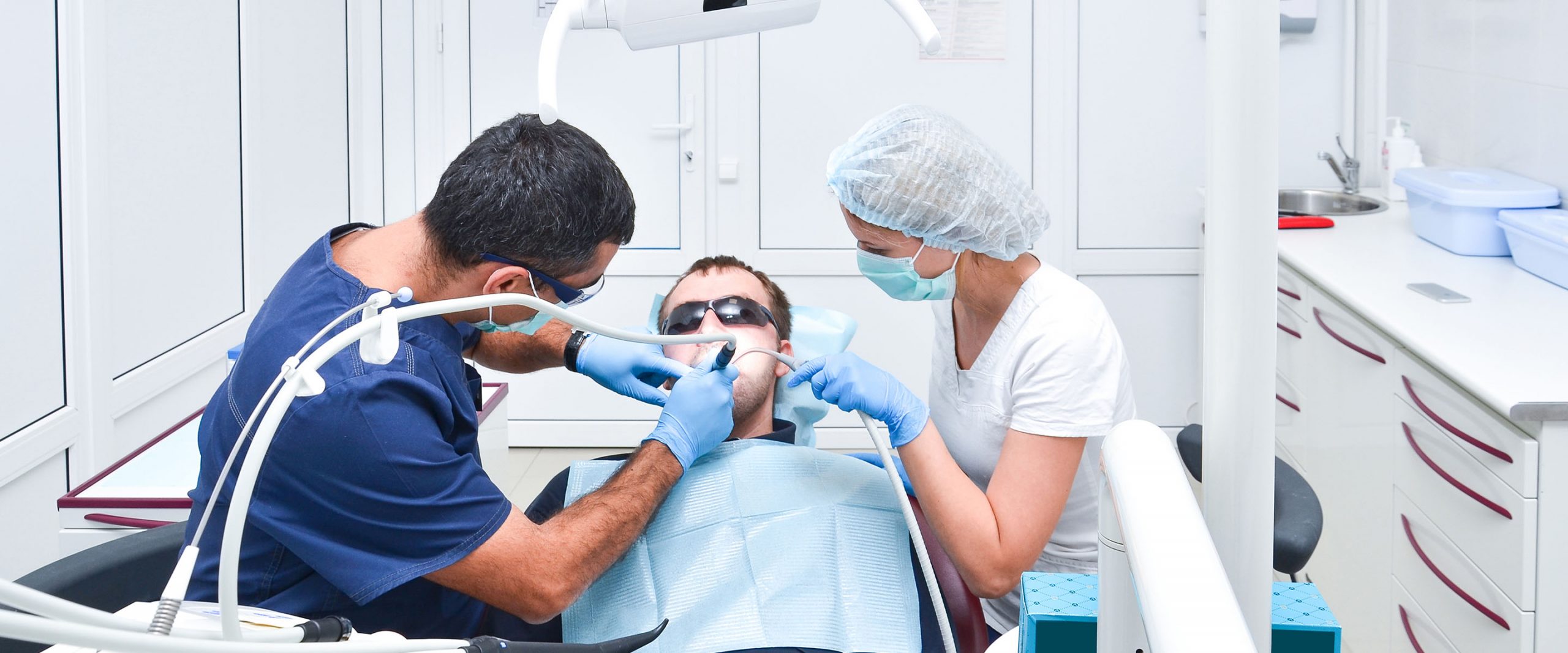
The text and images below are all based on my personal experience with dental implant restorations over the past 30 years.
Single units
Single unit abutment selection has been simplified by the “ti-base” family that most implant companies have adopted. These abutments have a retentive portion to which the laboratory manufactured prostheses is cemented extra-orally. The abutments come in different diameters and gingival cuff heights. The selection of the correct height and diameter should be done clinically. The problem with standardizing this very straightforward routine is illustrated in the x-ray image in fig. 1.

This patient consulted me with severe pain, redness and swelling around a freshly completed implant crown restoration. The history was as follows: An analog impression from implant level was taken and sent to the laboratory to fabricate a crown. The technician had no guidance as to where the implant neck was situated in relation to the soft tissues and bone. The shortest gingival cuff height (GH) was selected as this is esthetically the safest in order for a metal margin not to show at the gingival crest. On delivery of the implant crown, the dentist experienced difficulty “seating” the crown to the implant interface. The crown was placed with considerable resistance to achieve the required torque value. Impingement of the hard and soft tissues by the very acute emergence profile caused pressure to the bone, which will lead to necrosis and eventually marginal bone loss in that area. Selection of the correct abutment is required to avoid this complication.
Using an intraoral radiograph after implant placement or exposure (Fig. 2), the dentist may select the appropriate gingival cuff height for the abutment and guide the technician by providing the x-ray and/or the specification of the healing abutment. Most implant systems have synchronized shapes and contours of the surgical abutments and the prosthetic components.

Clinical example


Our challenge now, with these newer technologies, is to obtain a perfect cementation/bond between the restoration and the ti-base abutment. This is the area where most complications arise due to debonding or fracture of the zirconium base where the supporting thickness around the abutment is inadequately designed.
Multiple Units
Multiple implants are rarely parallel to make use of engaging components to implants placed at bone level. Usually placed below the soft tissue level, they are difficult to access with a mutual path of insertion. Non-engaging or “bridge” abutments are available to be used from implant level, but these can leave voids in the implant well, which with an ever-so-slight misfit will cause micro-leakage at the bone crest which could contribute to crestal bone loss.
It becomes complicated to obtain passivity and a path of insertion of the prosthesis when the implants are not placed parallel (Fig. 15)

In my opinion everything is simplified if an intermediate abutment is selected in multi-unit cases with the advantages listed as follows:
- Easy access to the prosthetic interface with ease of prosthetic procedures: impressions, placement
- Comfort of working and cleanliness of the access path with fluid-free interfaces
- Simplified path of insertion
- Passivity of fit
- Correction of alignment issues with angled abutments
- The muco-gingival tissue seems healthier than with a bone level interface
Some planning by the clinician is required, as these abutments should be selected before impressions or intra-oral scans are made to produce the final prosthesis. It is not accurate to connect these abutments on a cast made from an implant level impression and then fabricate the prosthesis.
Most implant systems have such intermediary abutments designed for the above advantages, mostly known as multi-base abutments, multi-unit abutments, convertible abutments, screw-retained abutments, conical abutments, universal abutments etc.
Clinical Example
Our patient illustrated in figures 16 &17, fractured the left central incisor and opted to have all four anterior teeth removed and replaced with implant-supported crowns.


Conclusions and clinical recommendations
1. In single tooth replacements, select an abutment which allows:
- Screw retention
- In laboratory cementation to a pre-fabricated abutment
- Digitally controlled fit to the retentive portion of the abutment (.stl file library)
- Which has the correct length to duplicate the surgical placed abutment profile
- Which does not apply pathologic pressure or compression of the surrounding tissue
- Places the cemented abutment interface away from the implant interface and surrounding bone
2. In multiple unit replacements or implant supported bridges:
- Select an intermediate abutment to fulfill the above requirements
- Select an intermediate abutment to eliminate parallelism and prosthesis passivity issues
- Select an intermediate abutment to bring the restorative interface easily accessible and clean
Learn more: Abutment Selection for Fixed Dental Prostheses
Acknowledgments
Dr. L.C. Swart, maxillofacial and oral surgeon, performed all the surgical procedures
Bobby Boshoff, OUTCAD, Digital Design Laboratory
Ian Robertson, Designer Dental Laboratory